
- 29 Jul, 2026
- Dr. Arun Rajeswaran
- No comment
MRI vs CT Scan: Understanding the Differences in Neuroimaging and Spine Care
In looking at conditions like recurrent headaches, back pain, or other changes related to brain function, doctors resort to sophisticated imaging methods to determine the root cause of the problem. In the debate of MRI Vs CT Scan, both techniques offer clear views of the body’s internal structures, but they differ in how they work and the medical conditions they are best suited to diagnose.
Key Technological Differences
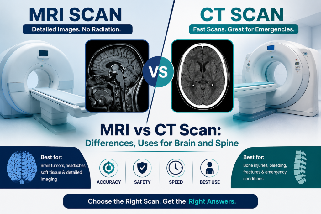
- CT Scan (X-Ray Based): Uses rotating X-ray beams coupled with computer processing to generate 3D cross-sectional images. Scans are rapid (typically 2 to 5 minutes) and widely accessible, making them the standard first choice in emergency medicine.
- MRI Scan (Magnetic & Radio Wave Based): Uses powerful magnetic fields and radiofrequency pulses to detail internal soft tissues without ionizing radiation. Scans take longer (15 to 45 minutes) and require patients to remain still inside a enclosed tube, but deliver higher soft-tissue contrast.
MRI Vs CT Scan in Neuroimaging & Spine Care
When CT Scans Are Preferred
- Acute Trauma & Fractures: Superior for evaluating bone detail, spinal fractures, bone spurs, and structural calcification.
- Emergency Hemorrhage Detection: Quickly identifies acute brain bleeding, intracranial trauma, or early hemorrhagic stroke.
- Patients with Metallic Implants: Safer alternative for patients with non-compatible pacemakers, deep-brain stimulators, or certain metallic fragments.
When MRI Scans Are Preferred
- Spinal Cord & Disc Pathologies: Best for evaluating herniated discs, spinal cord compression, pinched nerves, and sciatica.
- Soft-Tissue Brain Conditions: Preferred for detecting brain tumors, multiple sclerosis plaques, stroke ischemia, and subtle nerve lesions.
- Radiation-Free Safety: Safer choice for pediatric cases, pregnant patients, or individuals needing repeated follow-up imaging.
The Core Technology: How They Work
To understand when and why a specific scan is selected, it helps to understand the physical mechanisms behind these two powerhouse diagnostic imaging technologies.
Computed Tomography (CT Scan)
The CT Scan is basically a very advanced and quick process of conventional X-rays. Once you lay yourself in the round hole machine, a tube begins rotating around your body at an amazing speed. The machine fires multiple beams of X-rays through the selected area, and the rays are collected by the electronic detectors located on the other side.
The computer processing unit creates an overall image of the body from the multiple images taken. Since the CT Scan uses X-rays technology, it entails a dose of ionized radiation.
Magnetic Resonance Imaging (MRI Scan)
MRI is a procedure that does not involve any kind of radiation at all. Instead, it uses an extremely powerful magnetic field, combined with specific radiofrequency waves. Once you are placed inside the tube of the MRI machine, the extremely powerful magnetic field makes sure to align the hydrogen protons in your body’s water molecules.
However, the disruption of this alignment by the radiofrequency waves leads to the generation of signals from these protons, which in turn get converted into images by the computer.
Direct Architectural Differences
When choosing between these systems, medical teams weigh several operational factors:
- Primary Mechanism: A CT scan uses ionizing radiation (X-rays) to generate images, whereas an MRI relies entirely on strong magnetic fields and radio waves, leaving no radiation footprint.
- Scan Duration: CT scans are exceptionally fast, taking between 5 to 10 minutes from start to finish. An MRI is a much more drawn-out process, frequently lasting anywhere from 30 to over 60 minutes depending on the complexity of the sequences.
- Structural Visuals: CT scanning excels at capturing dense bone structures, acute internal bleeding, and calcified tissue. Conversely, an MRI offers unmatched clarity for soft tissue imaging, making it the premier choice for mapping fine organs and nerves.
- Patient Environment: The CT scanner is a shallow, spacious ring that operates completely silently. The MRI is a deep, enclosed tube that creates intense, rhythmic tapping and banging noises throughout the procedure, sometimes requiring ear protection.
MRI vs CT Scan for Brain Imaging
When looking into the complexities of the central nervous system, selecting between an mri vs ct scan for brain diagnostics comes down to a careful balance of clinical urgency and structural precision.
When is a Brain CT Scan Preferred?
Time is tissue when it comes to acute neurological trauma. A CT scan is the absolute gold standard in emergency settings. If a patient presents with sudden, severe neurological deficits or trauma, a CT scan can be completed in under five minutes. It excels at detecting:
- Acute intracranial hemorrhage: Spotting fresh blood from an aneurysm or stroke immediately.
- Skull fractures: Finding bone structural failures post-trauma.
- Calcified lesions: Detecting hardened, calcium-dense areas within brain tissue.
When is a Brain MRI Scan Preferred?
When an emergency is ruled out, or when fine structural resolution is required, an MRI is unmatched. Because it provides vastly superior soft tissue fidelity, an MRI allows neurosurgeons to see subtle variations in tissue density, micro-lesions, and delicate cellular structures that a CT completely misses. It is widely used to identify:
- Demyelinating conditions like Multiple Sclerosis (MS).
- Ischemic strokes in their very earliest hours.
- Subtle structural congenital defects.
MRI or CT Scan for Headache?
A highly frequent patient dilemma revolves around choosing an mri or ct scan for headache evaluation. If you suffer from standard migraines or occasional tension headaches with no warning signs, routine advanced imaging is rarely indicated.
However, if your headache is accompanied by “red flags”—such as a sudden “thunderclap” onset, fever, neck stiffness, confusion, or localized weakness—imaging becomes vital.
- Choose a CT Scan if: You experience a sudden, blinding headache that peaks within seconds. This pattern can signify a subarachnoid hemorrhage (bleeding around the brain), requiring immediate, rapid-fire intervention.
- Choose an MRI Scan if: Your headaches are chronic, progressively worsening, or accompanied by subtle neurological changes over weeks or months. An MRI provides the deep tissue fidelity required to rule out underlying structural anomalies, vascular malformations, or deep fluid collections.
Which Scan is Better for a Brain Tumor?
Whenever there is a suspicion of an intracranial mass lesion, a very important question comes to mind, what is better for brain tumor diagnostics? Even though CT is often ordered as a quick way of confirming the presence of the mass, MRI is unanimously recognized as a superior technique for brain tumor assessment.
With MRI imaging, the distinction between tumor tissue and normal brain tissue is excellent. Using contrast dyes (gadolinium, for example), MRI enables visualizing the exact borders of the tumor, the presence of internal blood vessels, and distinguishing tumor tissue from cystic fluids and edema.
In case you have an appointment with the neurosurgeon who will perform the complex craniotomy on the tumor, he needs high-resolution information about brain structures to operate around the eloquent areas that provide your speech, movement, and visual centers.
MRI Vs CT Scan for Spine Conditions
Descending from the cranium to the axial skeleton, when choosing between a ct scan and mri for the spine evaluation, one has the same diagnostic approach. It depends only on the necessity of examining either bone structure or the nerve tissue within.
Spine CT Scans
Since X-rays penetrate only denser tissues, a CT scan reveals an amazing resolution of bone structures called vertebrae. This technique can be used effectively in the diagnosis of complicated spine fractures, spinal instability, osteophytes, and serious bone damage seen in osteoarthritis or spinal stenosis.
Spine MRI Scans
Your spine is far more than just a column of bone; it houses the delicate spinal cord, exiting nerve roots, and shock-absorbing intervertebral discs. An MRI is the definitive imaging tool for mapping these neural structures. It is heavily relied upon to diagnose:
- Herniated or bulging discs: Seeing precisely how far a disc has protruded into the canal.
- Spinal cord compression: Finding regions where the cord is pinched or narrowed.
- Spinal infections or tumors: Highlighting inflammatory changes or neoplastic growths within the soft tissues of the canal.
Finding the Right Path for Your Neurological Health
Navigating brain and spine symptoms can feel overwhelming, but you do not have to make these diagnostic choices alone. Both CT scans and MRIs are vital, complementary assets in modern radiology.
If you are suffering from persistent headaches, debilitating back pain, or have questions regarding an upcoming brain or spine procedure, consult an expert. Visit Dr. Arun Rajeswaran’s official website to explore advanced treatment pathways and secure your path toward optimal neurological health.