
- 31 Jul, 2026
- Dr. Arun Rajeswaran
- No comment
Navigating the Diagnosis of Brain Tumors: Symptoms, Tests, and Next Steps
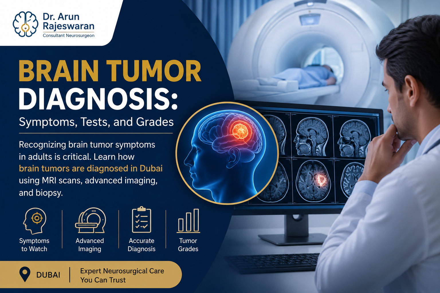
Brain Tumor Diagnosis: Learn how brain tumors are diagnosed in Dubai using MRI scans, advanced imaging, biopsy, and early symptom evaluation.
The prospect of undergoing a neurological examination may be extremely intimidating. The moment the patient or his family begins searching for an answer about an intracranial mass, it is important to learn more about diagnosing brain tumors.In the sphere of advanced neuro-oncology, it should be noted that a diagnosis of brain tumors does not involve a particular symptom or one particular procedure. It is a detailed multi-step puzzle gathered by an experienced team of specialists.If you want to diagnose a brain tumor in Dubai, the diagnostic process will involve the combination of high-level professional skills and modern neuroimaging techniques.
Recognising Brain Tumor Diagnosis Symptoms in Adults
Every diagnostic process is often initiated by the identification of minor changes in physical health. There is no consistency regarding the symptoms associated with brain tumors since they entirely depend on the position, the size, and the rate at which the tumor is developing in the skull.
The brain is tightly packed; when an abnormal tissue mass develops, it creates local pressure or interrupts electrical pathways. Some of the most common brain tumor symptoms in adults include:
- Progressive, Altered Headaches: This is often a deep, dull ache that feels noticeably worse in the early morning or when coughing, bending over, or straining.
- New-Onset Seizures: Experiencing a seizure for the very first time in adulthood is a major indicator that requires immediate investigation.
- Gradual Cognitive or Personality Shifts: Subtle changes in memory, difficulty concentrating, unexplained mood swings, or uncharacteristic behavior changes.
- Sensory and Motor Deficits: Progressive weakness, numbness, or loss of control in an arm or leg, often affecting just one side of the body.
- Vision and Balance Issues: Blurred vision, double vision, loss of peripheral sight, or persistent unsteadiness while walking.
The Initial Consultation and Neurological Assessment
A detailed examination is first performed before prescribing specific tests for brain tumors. In your first consultation with a neurosurgeon, you will be asked to provide your entire medical history in a chronological order followed by a physical examination that is non-invasive.
Your cranial nerves, which involve testing of your eye movements, facial symmetry, and pupil response, muscle strength, reflexes of the tendons and sensory perception are examined. If the physical findings suggest a localized structure problem within the central nervous system, further imaging procedures will immediately follow.
How Are Brain Tumors Diagnosed? The Role of Imaging
Brain tumor diagnosis relies on advanced brain imaging as the foundational cornerstone for detecting intracranial pathology. Modern diagnostic technologies allow specialists to look directly through bone and tissue to map anomalies with incredible geometric precision. These advanced imaging techniques help ensure accurate diagnosis and support timely treatment planning.
Magnetic Resonance Imaging (MRI Brain)
A brain MRI is the gold standard diagnostic imaging technique used to detect intracranial tumors. By making use of magnets and radio waves rather than radiation, an MRI gives the most detailed structural images of soft tissues.
For locating a suspected tumor, a contrast dye (gadolinium) is injected into the bloodstream. Contrast is used in areas where there is rapid uptake of the blood flow by the tumor, enabling the medical practitioner to get the precise outline of the tumor.
More sophisticated techniques, such as Magnetic Resonance Spectroscopy (MRS), are able to analyze the chemical makeup of the tumor, while Functional MRI (fMRI) analyzes critical areas of language and movement adjacent to the tumor site.
Computed Tomography (CT Scan)
Though an MRI may have better tissue contrast, the CT scan proves extremely useful in emergencies or preliminary diagnostics. The process consists of taking quick cross-sectional x-ray pictures which enable one to spot quickly any significant swelling, masses, hemorrhage, or problems with the adjacent cranial bones.
Confirming the Malignancy: Biopsy and Tumor Grading
While an advanced MRI can pinpoint a mass and suggest its likely identity, an imaging scan alone cannot provide a definitive cellular diagnosis. For accurate brain tumor diagnosis, the absolute confirmation of a brain tumor requires examining the tissue cells under a microscope. This step ensures the diagnosis is confirmed at the cellular level and helps guide the most appropriate treatment.
The Role of a Biopsy
Biopsy involves removing a tiny piece of the suspected tissue using a surgical process. A neurosurgeon could use this process when performing open surgery to remove the whole mass, which is referred to as craniotomy. In case the tumor is located in a very delicate area of the brain, then the neurosurgeon may conduct a needle biopsy.
With the help of computer navigation systems, accurate three-dimensional coordinates are used to guide a special needle to collect a tiny cell sample via a burr hole.
Tumor Grading and Pathology
Once the sample is collected, a neuropathologist analyzes the cells to determine the precise tumor type and assign a specific grade based on the World Health Organization (WHO) classification system:
- Grade 1 and 2 (Low-Grade): The cells look relatively normal under a microscope, grow slowly, and are less likely to spread aggressively into nearby healthy brain tissue.
- Grade 3 and 4 (High-Grade): The cells look highly abnormal, reproduce rapidly, and aggressively infiltrate surrounding functional brain structures, requiring rapid, multi-modal medical intervention.