
- 21 Oct, 2026
- Dr. Arun Rajeswaran
- No comment
Cranioplasty Surgery: Restoring the Skull After Brain Surgery
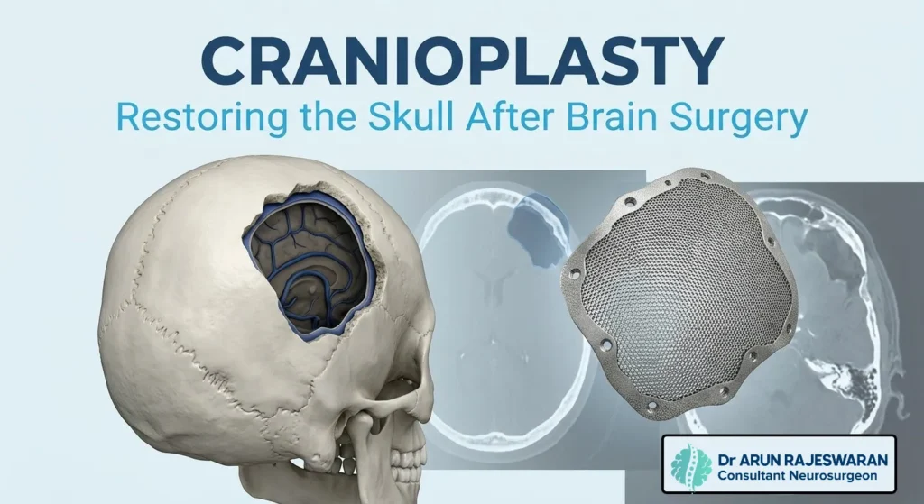
For patients who have undergone emergency brain surgery involving removal of part of the skull, the path to recovery often includes a second, carefully planned procedure to restore that missing bone. Cranioplasty surgery is that reconstructive step, and while it may sound purely cosmetic, it plays a significant role in both protection and neurological recovery.
In Cranioplasty Surgery, Why Part of the Skull Is Removed in the First Place?
Cranioplasty surgery almost always follows a prior procedure called decompressive craniectomy, an emergency surgery where a section of the skull is removed to relieve dangerous pressure inside the brain. This is typically performed after severe traumatic brain injury, a large stroke, or significant brain swelling from other causes, where the swelling brain needs room to expand without being compressed against a rigid, closed skull. The removed section of bone, sometimes called the bone flap, is either stored for potential future replacement or, in some cases, cannot be safely reused, requiring a custom implant later.
What Cranioplasty Surgery Involves
Skull reconstruction after brain surgery involves surgically placing material over the skull defect to restore its structure and protective function. Depending on the individual case, this can involve one of two general approaches.
| Approach | Description | When It Is Used |
|---|---|---|
| Bone flap replacement | The patient’s own previously removed bone is reattached. | When the original bone flap was preserved and remains viable. |
| Custom skull implant | A manufactured implant, often titanium or a medical-grade polymer, replaces the missing bone. | When the original bone was not preserved, is damaged, or has been resorbed. |
Titanium Skull Implant and Other Material Options in cranioplasty surgery
When a titanium skull implant or synthetic material is needed instead of the patient’s own bone, it is typically custom designed using detailed imaging of the patient’s specific skull defect. This custom approach ensures a precise fit and restores the natural skull contour as closely as possible. Titanium has long been a preferred material due to its strength and biocompatibility, though other materials such as certain medical grade plastics are also used depending on the specific case and surgeon’s recommendation.
Why Cranioplasty Surgery Often Waits
Cranioplasty is rarely performed immediately after the original decompressive craniectomy. There are important medical reasons for this delay.
- The brain and surrounding tissue need time to fully settle and reduce swelling before the skull is closed again
- Any wound healing or infection risk from the original surgery needs to resolve first
- Overall medical stability needs to be confirmed before undergoing a second procedure
The typical waiting period ranges from a few weeks to several months, depending on the patient’s recovery trajectory and the reason for the original craniectomy.
What Post-Craniotomy Recovery Looks Like Before Cranioplasty Surgery
During the interval between decompressive craniectomy and cranioplasty surgery, patients typically wear a protective helmet whenever out of bed, since the brain is unprotected by bone in the affected area during this period. This stage of post-craniotomy recovery often includes ongoing rehabilitation for any neurological deficits from the original injury, alongside careful monitoring of the skin and soft tissue overlying the skull defect.
Why Cranioplasty Matters Beyond Appearance
While restoring the skull’s normal contour does have a meaningful cosmetic benefit, neurosurgical reconstruction of the skull serves several functional purposes that go well beyond appearance.
- Physical protection, restoring a rigid barrier over the brain to prevent injury from everyday bumps or falls
- Neurological improvement, some patients experience measurable improvement in cognitive function and reduced headaches after cranioplasty, thought to relate to normalized pressure dynamics and cerebrospinal fluid flow
- Reduced risk of sunken skin flap syndrome, a condition where the skin over the defect sinks inward, sometimes causing neurological symptoms that can improve once the defect is repaired
What the Cranioplasty Surgery Itself Involves
Cranial defect repair surgery typically involves reopening the original surgical site, carefully preparing the edges of the existing skull defect, and securing either the preserved bone flap or the custom implant into place using small plates and screws designed to hold it securely while the surrounding tissue heals around it.
Recovery After Cranioplasty
Recovery from cranioplasty itself is generally more straightforward than the original emergency surgery, since it does not involve direct manipulation of brain tissue. Most patients spend a few days in the hospital for monitoring, followed by a gradual return to normal activity over the following weeks. Protective precautions around head injury are typically maintained for a period after surgery while the area fully heals and integrates.
Conclusion
Cranioplasty is the reconstructive procedure that restores the skull following decompressive craniectomy, using either the patient’s original bone flap or a custom titanium or synthetic implant depending on the individual case. Beyond restoring normal appearance, this cranial defect repair serves genuine functional purposes, protecting the brain from injury, and in some patients supporting measurable neurological improvement by normalizing pressure dynamics that were disrupted while the skull defect remained open. Understanding this second stage of recovery helps patients and families see it as a meaningful part of the healing process rather than a purely cosmetic afterthought.