
- 31 Jul, 2026
- Dr. Arun Rajeswaran
- No comment
The Ultimate Guide to Spine Imaging: When Do You Need a Scan for Back Pain?
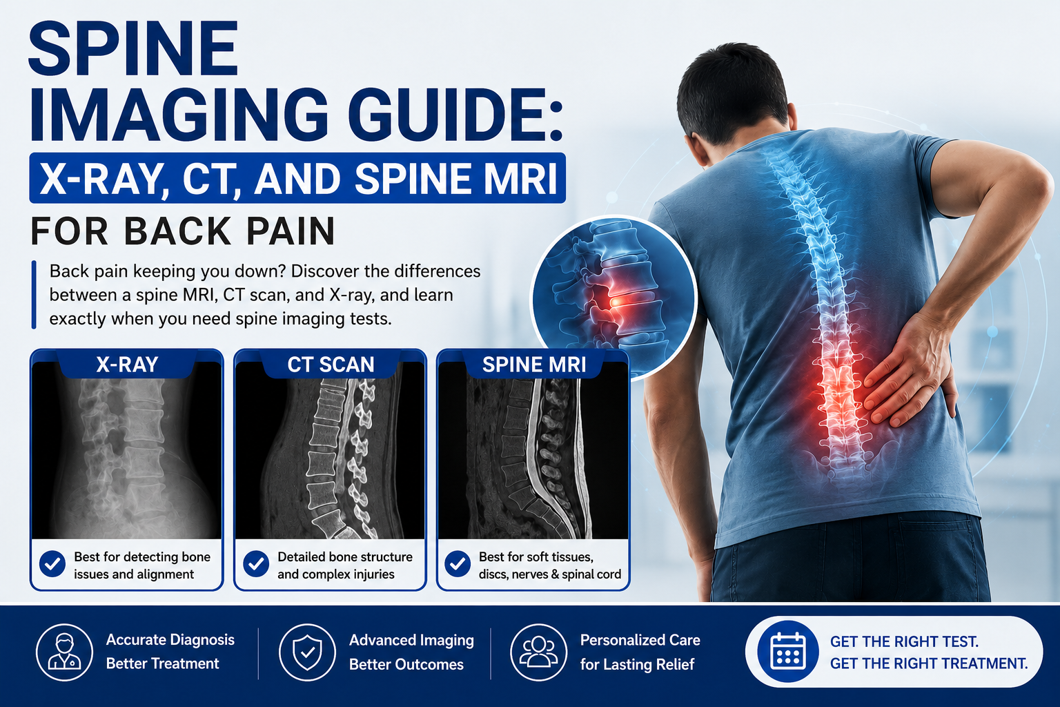
Back pain keeping you down? Discover the differences between a spine MRI, CT scan, and X-ray, and learn exactly when you need spine imaging tests.
Most people suffer from either back pain or neck pain at some stage in their lives. Often, such injuries heal on their own without any intervention within a couple of weeks of rest, mild stretches, and careful treatment. But in the case where spinal pain becomes chronic or serious, or even associated with neurologic changes, the healthcare provider needs to look beyond and find out the structural problem behind it.
Spine imaging is considered the main guideline for treating spinal disorders. Whether you need to identify the source of your painful herniated disc or follow the progress of your degenerative spine disorder, the advanced diagnostic methods make the decision-making process much easier.
Deciding whether to choose a spine MRI, a CT scan or an ordinary X-ray may be quite a difficult task. The knowledge about what each procedure involves may help you navigate through the options.
When Do You Need Spine Imaging?
A highly common misconception among patients is that a scan is required the moment back pain begins. In clinical practice, routine imaging is rarely indicated for acute, uncomplicated lower back pain during the first four to six weeks.
Physicians lean on a specific set of clinical indicators—often referred to as “red flags”—to determine exactly when you need spine imaging. Diagnostic scans become essential if you experience:
- Progressive Neurological Deficits: Sudden or worsening weakness in your legs or arms, difficulty lifting your foot while walking, or clumsy hand movements.
- Radicular Pain: Sharp, shooting pain or electrical sensations that travel down your buttocks into your leg (sciatica) or radiate down your shoulder into your arm, pointing toward potential nerve compression.
- Persistent Numbness or Tingling: Chronic loss of sensation or a permanent “pins and needles” feeling in your extremities.
- Trauma or Impact: Back pain that originates immediately following a motor vehicle accident, a severe fall, or a sports-related impact.
- Constitutional Symptoms: Unexplained weight loss, fever, or a history of oncology conditions alongside new, unremitting back pain that feels worse when lying down at night.
- Bowel or Bladder Dysfunction: Sudden inability to pass urine or an unexpected loss of bowel control. This is a medical emergency known as Cauda Equina Syndrome, requiring immediate imaging and surgical intervention.
Comparing Your Options: Spine X-Ray vs MRI vs CT Scan
Various modalities of imaging are tailored for visualization of different tissues within the human body. The type of modality that would be chosen by a specialist depends on what part of your vertebrae – the dense bone structure or the fragile neurological tissues – is under study.
1. Spine X-Ray: The Initial Structural Baseline
A standard spine X-ray is often the first visual step in an outpatient evaluation. It uses a very low dose of radiation to capture flat, two-dimensional images of the bones.
- What it sees best: Spinal alignment, severe joint degeneration, advanced osteoarthritis, bone spurs, and structural instability like spondylolisthesis (where one vertebra slips over another).
- What it misses entirely: X-rays cannot see soft tissues. They offer zero visibility into the health of your intervertebral discs, the spinal cord, or individual nerve roots.
2. Spine MRI: The Gold Standard for Soft Tissue
MRI imaging for back pain is unmatched if there are any problems related to either the nerves or the discs. Instead of using radiation like CT scans, MRI scanning uses radiofrequency waves and powerful magnets to get detailed images of the spinal column’s soft tissue areas.
In an MRI brain or spine scan, the neurosurgeon can determine the amount of fluid left within a disc, define the borders of the herniated disc, and see whether the passage of the spinal cord is constricted.
3. CT Scan for Spine: High-Definition Bone Mapping
Computed Tomography (CT) scan uses highly specialized X-ray equipment along with advanced computer technology to create detailed cross sections of the body.
Though MRI works well with soft tissues, CT scan of the spine remains the number one choice in order to analyze the details of bones. This test is frequently performed to diagnose complicated fractures of the spine in case of injury, check spinal fusion, or changes in bone density in case the patient cannot go through MRI because of a pacemaker or metal devices.
Common Spinal Disorders Identified Through Imaging
Advanced diagnostics allow medical teams to accurately categorize and name the underlying structural issues driving your discomfort. The most common structural abnormalities discovered include:
Herniated and Bulging Discs
The intervertebral discs serve as rubber-like cushions separating each vertebra in your spine. With age or because of any kind of sudden trauma, the hard outer layer of this disc becomes worn out. The inner layer of jelly-like substance starts protruding or even ruptures completely, leading to pressure on the surrounding structures.
Degenerative Spine Disease
This is a general term for the process whereby the spine gets worn out due to the aging process. Due to the loss of fluid in the discs, they tend to become flattened. As a result, there is little space left that causes excessive strain to the facet joints in the spine which leads to osteophyte formation and inflammation.
Spinal Stenosis
If the spinal canal and the openings where the nerves branch off become compressed because of bony overgrowths or bulging discs, it leads to squeezing of the spinal cord and nerves. The problem is termed as spinal stenosis, and its symptoms are cramping, numbness, and severe pain in the legs during standing or walking.
Taking Control of Your Spinal Wellness
Having chronic pain in your back or neck will have a major effect on your quality of life, but being able to get a proper diagnosis of your condition will help you regain some of your mobility. Proper imaging testing will require sound clinical judgment depending on your physical symptoms.
If you have chronic back pain or radiating nerve pain, then getting proper guidance in having your spine imaged is very important.