
Blog
Hydrocephalus Treatment: A Neurosurgeon’s Guide to Shunts, Endoscopic Surgery, and Managing CSF Pressure
- 29 May, 2026
- Dr. Arun Rajeswaran
- No comment
Hydrocephalus Treatment: A Neurosurgeon’s Guide to Shunts, Endoscopic Surgery, and Managing CSF Pressure
Most patients have never heard the word hydrocephalus until the day it applies to them.
“Water on the brain” is the phrase people often use. It sounds frightening. The reality is more manageable than those words suggest, and understanding what is actually happening inside the skull makes the whole situation far less overwhelming.
Hydrocephalus is a build-up of cerebrospinal fluid inside the brain’s ventricles the fluid-filled spaces deep within the brain. When fluid accumulates faster than it drains, pressure rises. That pressure causes the symptoms patients feel. Left untreated, it causes damage.
The good news is that hydrocephalus is treatable. The right surgical procedure, chosen carefully for the right patient, restores normal fluid drainage, relieves pressure, and gives most people their quality of life back. Getting there requires an accurate diagnosis, a clear understanding of what type of hydrocephalus is present, and the right operation.
This article walks through all of that.
Every day, the brain produces around 500ml of cerebrospinal fluid. This fluid circulates through the ventricular system, flows around the brain and spinal cord, and gets gradually reabsorbed into the bloodstream.
In a healthy system, production and absorption stay in balance. Pressure remains stable.
In hydrocephalus, that balance breaks down. Either too much fluid is produced, not enough is absorbed, or normal flow is blocked somewhere along the route. The ventricles enlarge to accommodate the fluid that cannot drain. Pressure climbs. Brain tissue gets compressed.
How quickly this happens determines how symptoms present. In acute hydrocephalus, pressure rises fast and symptoms come on hard. In the slower forms, particularly normal pressure hydrocephalus in older adults, things shift gradually over months or years. Patients often assume it is just ageing.
It is not always just ageing.
Two Types, Two very different problems
Not all hydrocephalus is the same. Getting the type right is what leads to the correct operation.
Obstructive hydrocephalus occurs when something physically blocks the flow of CSF within the ventricular system. The cerebral aqueduct, a narrow channel connecting the third and fourth ventricles, is the most common site. Tumours, cysts, old scarring from infection or bleeding, and certain congenital abnormalities can all cause this blockage.
Communicating hydrocephalus is different. Here, fluid moves freely between the ventricles, but it cannot be absorbed properly once it reaches the outer surfaces of the brain. The drainage point, the arachnoid granulations, is where the problem sits. Causes include previous meningitis, subarachnoid haemorrhage, and inflammatory conditions.
Normal pressure hydrocephalus (NPH) is a specific variant of communicating hydrocephalus. It mainly affects people over 60. The ventricles are clearly enlarged on imaging, but measured CSF pressure sometimes appears within the normal range. That combination confuses the picture and delays diagnosis more than it should.
NPH has a recognisable clinical pattern: a shuffling, wide-based gait, urinary urgency or incontinence, and a slowing of cognition. All three together are classic pictures. Gait problems usually come first. Families often notice before the patient does.
More on NPH below, because it is commonly missed and commonly treatable.
How Hydrocephalus Is Diagnosed
Diagnosis starts with a detailed conversation. How long have symptoms been present? Did they come on suddenly or drift in gradually? Are headaches worse in the morning? Has walking changed? Has memory or thinking changed?
A neurological examination follows. Then imaging.
MRI is the primary investigation. It shows the size of the ventricles, identifies any blockage or structural cause, and shows how the surrounding brain tissue looks. CT scanning is used in urgent situations where speed matters more than detail.
For NPH specifically, diagnosis involves extra steps. A large-volume lumbar puncture, removing 30–50ml of CSF, is performed to test whether symptoms improve when pressure is briefly reduced. If a patient walks noticeably better after the tap, that predicts a good response to shunting. Continuous intracranial pressure monitoring, done over 24 hours or longer, can detect abnormal pressure waves that a single lumbar puncture misses.
These investigations matter. The aim is not just to confirm hydrocephalus but to predict which patients will genuinely benefit from surgery.
VP Shunt Surgery
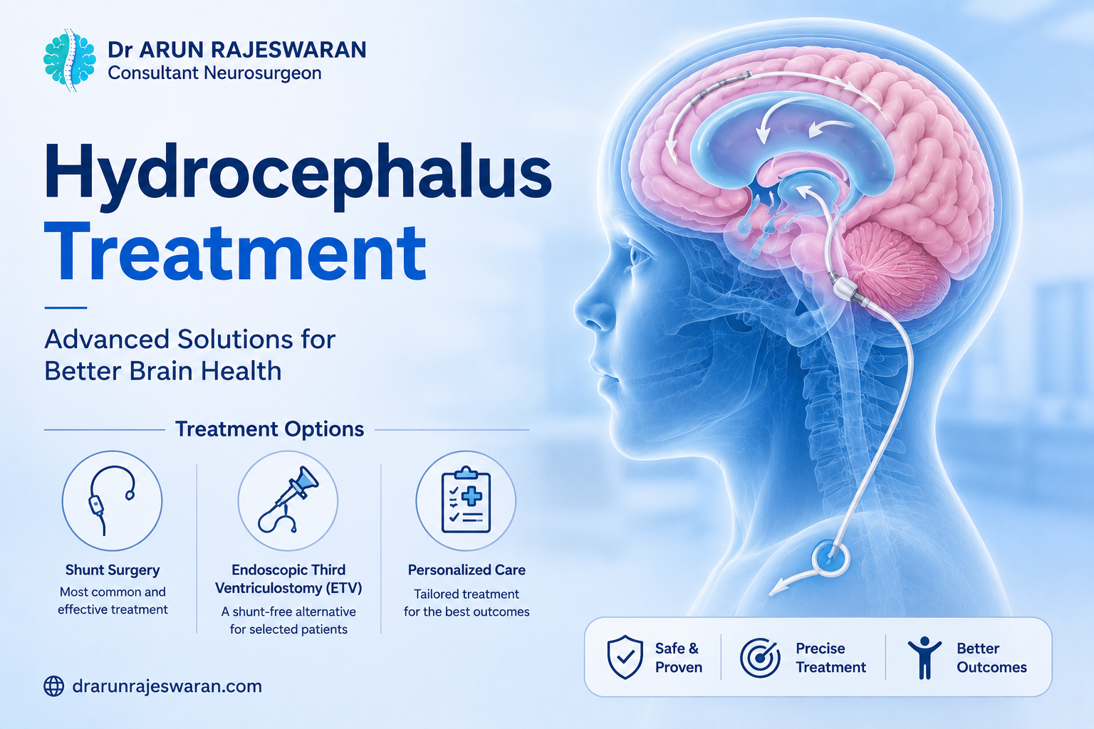
A ventriculoperitoneal shunt, known as a VP shunt, is the most widely used surgical treatment for hydrocephalus globally. It works by creating an alternative drainage route for CSF, bypassing whatever is stopping it from draining normally.
A thin, flexible catheter is placed inside one of the lateral ventricles of the brain. This connects to a pressure-sensitive valve, which sits just beneath the scalp. A second catheter runs from the valve down under the skin to the peritoneal cavity, the space inside the abdomen, where CSF drains and gets absorbed.
Surgery is done under general anaesthesia. Two small incisions are made, one on the scalp and one on the abdomen, and the catheter is passed under the skin between them. In experienced hands, the procedure takes around one to two hours. Most patients go home within a few days.
The valve is what controls the drainage. It opens when pressure exceeds a set threshold, lets CSF through, and closes when pressure returns to normal. Programmable valves allow that threshold to be adjusted non-invasively after surgery using a small external device. No return to the operating room is needed. This flexibility is particularly useful in NPH, where finding the right drainage setting takes some trial and adjustment.
VP shunts work for both communicating and obstructive hydrocephalus. They are the standard treatment for normal pressure hydrocephalus.
When Shunts Develop Problems
A shunt is a long-term implanted device. Devices can malfunction. Patients should know what to watch for, not to feel anxious about it, but because early recognition means faster treatment.
Blockage is the most common problem. The ventricular catheter can get clogged. The abdominal catheter can kink or shift position. When a shunt blocks, hydrocephalus returns. Headache is usually the first sign, often pressure-like, worse in the mornings. Nausea, drowsiness, and visual disturbance follow.
Infection affects roughly 5–8% of shunt procedures. It tends to present within the first few months, with fever, headache, and tenderness along the shunt tract. Infected hardware usually needs to come out. The patient is managed with antibiotics and a temporary external drain while the infection clears, then a new shunt is placed.
Over-drainage is less obvious but worth knowing about. If the valve setting allows too much fluid to drain, pressure drops lower than it should. Patients feel worse when upright and better lying flat. Imaging may show small fluid collections around the brain. Adjusting the valve setting usually resolves this.
Shunt revision surgery, to replace or reposition a blocked or failing component, is a routine part of living with a shunt over many years. It is not a complication in the alarming sense. It is planned, manageable, and usually straightforward.
Endoscopic Third Ventriculostomy (ETV)
ETV is a different approach entirely. Rather than inserting a drainage device, it uses the brain’s own anatomy to create a new pathway.
A small endoscope, a thin tube with a camera and light, is passed through a burr hole in the skull and guided into the third ventricle. A small opening is made in the floor of the ventricle. CSF can then bypass the obstruction and flow directly into the subarachnoid space, where it absorbs naturally.
The appeal is clear. If it works, there is no hardware at all. No shunt to block, no valve to fail, no infection risk from an implanted device.
ETV works best for obstructive hydrocephalus where there is a clear anatomical blockage and the subarachnoid space is in good condition. Aqueduct stenosis is the ideal scenario. Success rates are high in adults and older children with this presentation.
It is less suitable for communicating hydrocephalus, where the absorption pathway itself is the problem. Creating an opening in the ventricular floor does not fix impaired absorption. And in patients who have had previous infection, bleeding, or surgery, subarachnoid scarring can reduce how well the new pathway works.
ETV is done under general anaesthesia, typically in under an hour. Recovery tends to be quicker than shunt surgery.
The honest caveat is that ETV can fail, particularly in the first few months after surgery. A small proportion of patients return with symptoms and need a shunt. Patient selection is what makes ETV successful, not the procedure in isolation.
Emergency Drainage: The External Ventricular Drain
When hydrocephalus is acute and pressure is rising dangerously fast, there is no time to plan a shunt or ETV. An external ventricular drain buys time.
A catheter is placed directly into the lateral ventricle and connected to an external collection bag. CSF drains out, pressure drops, and the patient is stabilised. It is a temporary measure, not a long-term solution. Infection risk increases with every day the drain is in place, and it requires close monitoring in a hospital setting.
Once the immediate crisis is managed and the cause addressed, a definitive procedure follows.
Normal Pressure Hydrocephalus: The Treatable Condition That Gets Missed
NPH deserves its own section because of how often it goes unrecognised.
The three symptoms, gait disturbance, urinary urgency, and cognitive slowing, are each easy to dismiss on their own. Gait problems get attributed to arthritis or Parkinson’s. Bladder issues get put down to age. Memory slowly gets called dementia. Individually, that seems plausible. Together, they form a picture that should prompt a neurology or neurosurgery referral.
The gait in NPH is specific. Short steps. Wide stance. A tendency to shuffle. Difficulty lifting the feet off the floor. Patients describe it as their feet feeling stuck. It is different from Parkinson’s, and an experienced clinician can usually tell.
Shunting in well-selected NPH patients can produce significant improvement. Gait response is the most reliable. Cognitive improvement varies. Bladder control tends to improve less consistently. The key word is well-selected. Not every patient with enlarged ventricles has NPH, and not every patient with NPH responds to shunting. The tap test, careful imaging analysis, and sometimes longer-term pressure monitoring are what separate the patients who will benefit from those who will not.
One point on timing: the earlier NPH is treated, the better. Patients who have been symptomatic for several years, or who have significant brain atrophy on imaging, tend to see less recovery than those treated in the earlier stages. If this picture sounds familiar for someone you know, it is worth pursuing sooner rather than later.
Children and Hydrocephalus
This article is primarily about adults, but hydrocephalus affects all ages. The presentation differs.
In infants, the skull is still flexible because the sutures have not yet fused. Excess fluid causes the head to enlarge rather than pressure to build in the same way. A head circumference growing too fast, a bulging fontanelle, eyes that look downward, and unexplained irritability are the key signs. In older children, once the skull is fused, the symptoms look more like adult hydrocephalus: headache, nausea, vomiting, and neurological changes.
Treatment principles are the same. Shunting and ETV are both used. Surgical technique is adapted to the child’s size, and valve selection is particularly important in growing children.
How I Approach Hydrocephalus in Practice
When a patient with hydrocephalus comes to my clinic, the first priority is establishing what type it is and what is driving it. That question shapes every decision that follows.
The clinical history and examination come before anything else. What are the symptoms? When did they start? How have they progressed? A detailed neurological examination follows, assessing gait, cognition, reflexes, and coordination. Then imaging is reviewed carefully, not just to confirm hydrocephalus is present but to understand the underlying cause.
For obstructive cases with anatomy suitable for neuroendoscopy, ETV is usually the first choice. No hardware, faster recovery, and if it works, no long-term device to manage. For communicating hydrocephalus, NPH, and cases where ETV is not appropriate, a VP shunt with a programmable valve is what I use most often.
After surgery, follow-up is structured. Shunt settings are adjusted based on how the patient responds. Imaging is repeated to confirm the ventricles have decompressed. Patients and families are educated about the signs of shunt malfunction before they leave hospital.
Most patients with a well-functioning shunt go on to live completely normally. The shunt fades into the background. They stop thinking about it. That is the goal.
What to Expect After Surgery
Recovery and outcomes depend on the underlying cause, how long the patient was symptomatic before treatment, and whether any lasting pressure damage occurred.
For obstructive hydrocephalus treated with ETV, or pressure from a resolved haemorrhage treated with shunting, outcomes are usually good. Symptoms resolve, and long-term prognosis is strong.
For NPH, gait recovery is the most predictable result. Cognition improves in many patients but not all. Bladder symptoms are variable. Realistic expectations before surgery are part of the conversation I always have.
For hydrocephalus linked to malignant tumours or severe brain injury, prognosis is tied to the underlying condition as much as to the fluid dynamics. Shunting controls pressure. That is not a small thing. Preventing continued pressure damage to brain tissue has a real impact on quality of life, even when complete recovery is not possible.
When to Get a Specialist Opinion
If you have been told you or a family member has hydrocephalus and have not yet seen a neurosurgeon, that appointment should happen soon.
If you have an existing shunt and new symptoms have appeared, do not wait to see how things go. A worsening headache, drowsiness, vomiting, or visual changes in someone with a shunt needs prompt assessment.
If you have a parent or older relative showing the triad of shuffling gait, urinary urgency, and cognitive slowing, bring it to a doctor’s attention. NPH is underdiagnosed, and treatment delays reduce the chance of good recovery.
Hydrocephalus is serious. It is also well-managed with the right approach. If you have questions or want an evaluation in Dubai, I am here.
Frequently Asked Questions
It depends on the type. For obstructive hydrocephalus, where the CSF flow is physically blocked, endoscopic third ventriculostomy is often the best first option because it avoids an implant entirely. For communicating hydrocephalus and normal pressure hydrocephalus, a VP shunt is the standard and reliable treatment. Both approaches work by restoring normal drainage. The choice between them comes from carefully evaluating the cause, the patient’s anatomy, and their clinical history. There is no single answer that works for every case.
It is a routine neurosurgical procedure with a good track record in experienced hands. Like any operation, it carries risks: infection, bleeding, and the possibility of hardware problems over time. The complication most patients encounter over years of living with a shunt is blockage or malfunction requiring revision surgery. That said, the alternative, leaving elevated intracranial pressure untreated, causes progressive neurological damage. For the right patient, the benefits of shunting far outweigh the risks. Most people recover well and return to normal life within a few weeks.
The symptoms of shunt failure are essentially the symptoms of hydrocephalus coming back. Headache is usually first, often pressure-like and worse in the mornings. Nausea, drowsiness, and visual disturbance follow in more significant failure. In patients with NPH who were doing well after shunting, a gradual return of gait problems or cognitive slowing can also signal that the shunt needs attention. Any of these symptoms in a patient with a shunt should prompt a prompt medical review. It is not a situation to monitor at home.
In most cases, no. Medications like acetazolamide can temporarily reduce CSF production and are sometimes used as a short-term bridge, but they are not a lasting solution. Serial lumbar punctures can provide temporary symptom relief in NPH and are used diagnostically, but the effect does not last. There is no medication that permanently corrects the underlying drainage failure. For sustained relief of intracranial pressure, surgery is needed. The only real question is which surgical approach suits the individual patient best.
NPH mainly affects adults over 60. Despite the name, the ventricles are enlarged and damage from pressure is occurring, even though measured CSF pressure sometimes falls within the normal range. The classic presentation is three things appearing together: a shuffling, wide-based gait where the feet seem stuck to the floor; urinary urgency or incontinence; and a slowing of thinking and memory. Each symptom alone might suggest something else. Together, they point to NPH. This matters because NPH is one of the few reversible causes of dementia-like symptoms in older adults, and appropriate patients can improve substantially with VP shunt surgery.