
Blog
Causes of Balance Problems in Adults: Brain, Spine, and Inner Ear How to Tell the Difference
- 09 Jul, 2026
- Dr. Arun Rajeswaran
- No comment
Causes of Balance Problems in Adults: Brain, Spine, and Inner Ear How to Tell the Difference
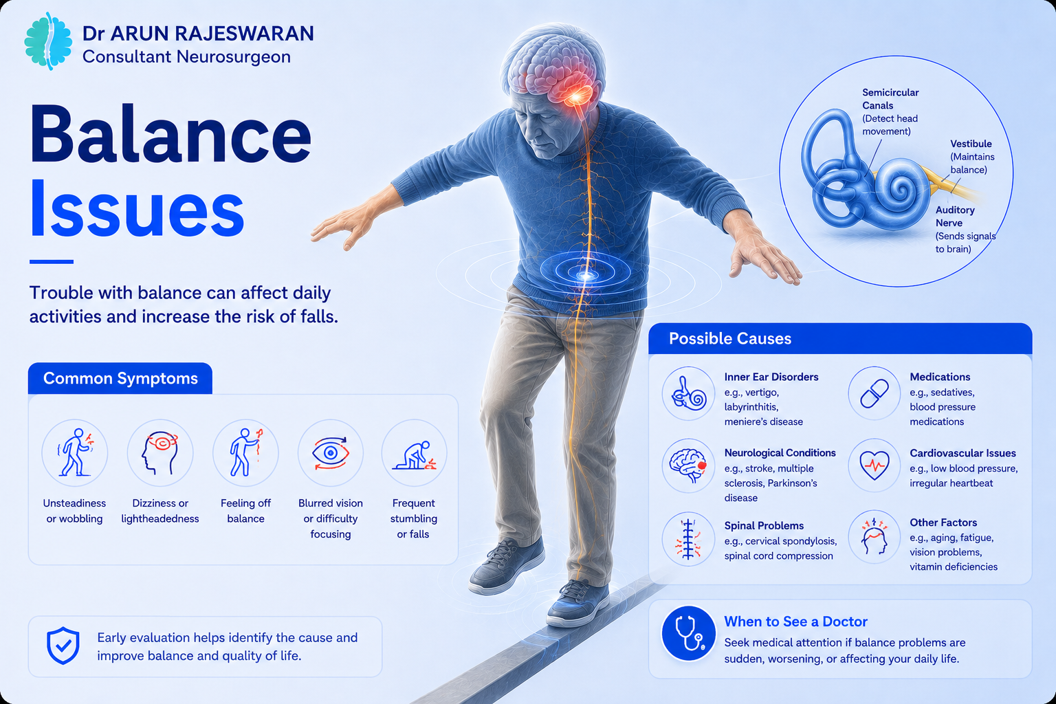
Balance problems in adults are frequently attributed to the inner ear or to ageing. In many cases that is correct. But balance is controlled by three systems working together: the inner ear, the eyes, and the brain and spinal cord and when the brain or spinal cord is the source, the cause is more serious and the assessment is different.
Understanding which system is responsible for unsteadiness guides both the investigations needed and the specialist required.
Vertigo vs Imbalance: A Critical Distinction
Feature | Vertigo (Spinning Sensation) | Imbalance (Unsteadiness) |
|---|---|---|
Symptom | Illusion of movement room spinning or self-spinning | Difficulty maintaining stable posture or gait; unsteady walking |
Inner ear origin | Common BPPV, vestibular neuritis, Meniere’s disease | Less typical for inner ear in isolation |
Brain origin | Possible cerebellar or brainstem lesion | Common cerebellum, spinal cord, or proprioception pathway |
Position triggered | Often BPPV strongly triggered by head position change | Not typically position-dependent |
Duration | Seconds to minutes (BPPV); hours to days (Meniere’s) | Often persistent; not episodic |
Nausea/vomiting | Common | May be present but less prominent |
Key red flag | Vertigo + sudden severe headache / diplopia / dysarthria | Progressive worsening over weeks; unsteady gait |
Inner Ear Causes
Benign Paroxysmal Positional Vertigo (BPPV)
The most common cause of episodic vertigo. Tiny calcium crystals dislodge within the semicircular canals, causing brief but intense spinning with specific head movements rolling over in bed, looking upward, bending forward. Each episode lasts less than a minute. Treated with canalith repositioning manoeuvres (Epley manoeuvre).
Vestibular Neuritis
Sudden-onset severe vertigo, usually after a viral illness, lasting days. Accompanied by nausea and vomiting. No hearing loss. Gradually improves as the brain compensates, but unsteadiness may persist for weeks.
Meniere’s Disease
Episodes of severe vertigo lasting minutes to hours, combined with fluctuating low-frequency hearing loss, tinnitus, and a sense of fullness in the ear. Managed medically by an ENT specialist.
Brain Causes
Cerebellar Disorders
The cerebellum at the back of the brain coordinates movement and balance. Damage to the cerebellum produces a characteristic unsteady, wide-based gait called cerebellar ataxia. Walking looks uncoordinated. Fine hand movements become clumsy. Speech may sound slurred.
Causes include stroke in the cerebellar circulation, cerebellar tumours, multiple sclerosis, and alcohol-related cerebellar degeneration.
Acoustic Neuroma (Vestibular Schwannoma)
A benign tumour growing on the vestibular nerve, the nerve connecting the inner ear to the brain. Produces gradual one-sided hearing loss, tinnitus, and unsteadiness. Grows slowly but can eventually compress the brainstem if untreated. Diagnosed on MRI.
Brainstem Disorders
The brainstem coordinates many of the signals involved in balance. Stroke, tumours, and demyelinating lesions in the brainstem produce balance problems that are often accompanied by double vision, facial numbness, swallowing difficulty, or limb weakness features that distinguish brainstem causes from inner ear causes.
Spinal Cord Causes
Cervical Myelopathy
Compression of the cervical spinal cord narrows the signals travelling to and from the brain, disrupting proprioception the body’s sense of limb position. Patients describe a sense of unsteadiness when walking, difficulty on uneven surfaces, and a feeling that the ground is not where they expect it to be. Leg stiffness develops. In later stages, the gait becomes narrow and cautious.
Cervical myelopathy is one of the most commonly missed diagnoses in adults with progressive gait problems. It looks like ageing. It is a treatable spinal cord condition.
Normal Pressure Hydrocephalus
In older adults, the combination of a shuffling wide-based gait, urinary urgency, and cognitive slowing constitutes the classic triad of normal pressure hydrocephalus. The gait is distinctive short steps, feet barely lifting from the floor, difficulty initiating walking. If this combination is present, neurosurgical assessment is appropriate.
When an MRI Becomes Necessary
Feature | MRI Indicated? | Urgency |
|---|---|---|
Sudden vertigo + headache, diplopia, or dysarthria | Yes urgent | Emergency (possible posterior fossa stroke) |
Progressive unsteadiness over weeks without resolution | Yes | Within days |
Unsteady gait + leg stiffness or hand clumsiness | Yes MRI spine | Urgent |
One-sided hearing loss + tinnitus + balance problems | Yes MRI brain | Within 1–2 weeks |
Classic BPPV resolving with Epley manoeuvre | Usually not required | As clinically guided |
Older adult with NPH triad (gait, bladder, cognition) | Yes MRI brain | Within 1–2 weeks |
Diagnosis and Tests
- Clinical neurological and vestibular examination: identifies gait type, nystagmus (involuntary eye movement), Romberg sign, and coordination testing
- MRI Brain: identifies cerebellar, brainstem, or acoustic neuroma lesions
- MRI Cervical Spine: identifies spinal cord compression causing gait disturbance
- Audiometry and vestibular function testing: assesses inner ear function
- Dix-Hallpike test: confirms BPPV at the bedside without imaging
When to See a Neurosurgeon
Neurosurgical assessment is appropriate when balance problems are linked to a structural brain or spine cause: an acoustic neuroma, a cerebellar tumour, brainstem compression, cervical myelopathy requiring decompression, or normal pressure hydrocephalus requiring shunting. Inner ear causes are managed by ENT. Mixed presentations may require both specialties.
Conclusion
Balance problems in adults have a broad range of causes. The distinction between inner ear, brain, and spinal cord causes determines the investigation pathway and the treating specialist. Progressive, non-episodic balance problems in an adult warrant neurological assessment rather than assumption that the cause is vestibular.
If you are experiencing persistent neurological symptoms, consult Dr. Arun for a detailed evaluation and personalised treatment plan.
Frequently Asked Questions
Sudden balance problems in adults require urgent assessment. Posterior fossa stroke affecting the cerebellum or brainstem produces sudden severe vertigo, unsteadiness, nausea, and vomiting, often with double vision, facial numbness, or limb weakness. This is a medical emergency. Vestibular neuritis also causes sudden onset but is not accompanied by other neurological features. BPPV causes sudden brief spinning triggered by head position change.
Yes. Tumours in the cerebellum or brainstem produce balance disturbance and unsteady gait. Acoustic neuromas on the vestibular nerve cause progressive one-sided hearing loss, tinnitus, and unsteadiness. Tumours in the fourth ventricle can cause hydrocephalus with gait disturbance. Balance problems that are progressive over weeks without fluctuation or position-dependence warrant brain MRI.
Vertigo is the false sensation of movement, the room spinning or the patient spinning. It is an inner ear or brainstem symptom. Balance disorder is difficulty maintaining stable posture or walking without falling. It reflects a broader set of causes including the cerebellum, spinal cord, and proprioceptive pathways. Both can co-exist, but their distinction guides the investigation and specialist referral.
Yes. Cervical myelopathy compression of the spinal cord in the neck disrupts the signals controlling limb position sense (proprioception), producing a characteristic unsteady, wide-based gait. Patients feel uncertain on their feet, particularly on uneven ground or in the dark. This is distinct from vestibular-related balance problems and is diagnosed on cervical spine MRI.
Bedside tests including Romberg test, tandem gait, and Dix-Hallpike test assess the vestibular and cerebellar systems. MRI of the brain examines the cerebellum, brainstem, and posterior fossa. MRI of the cervical spine identifies spinal cord compression. Audiometry and vestibular function tests assess inner ear function. Which tests are ordered depends on the clinical presentation.