
Blog
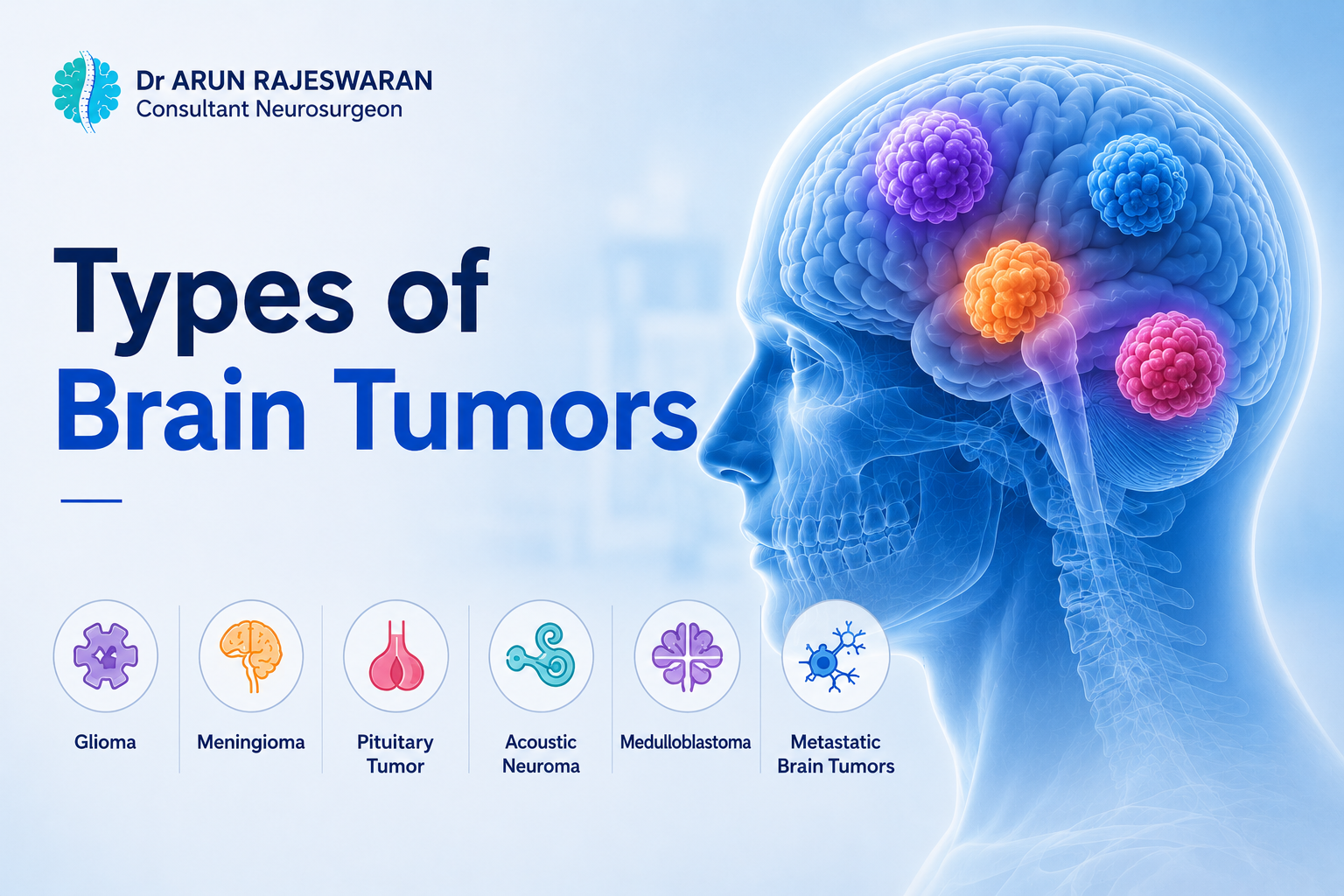
Types of Brain Tumors: A Clear Guide from a Neurosurgeon
- 29 May, 2026
- Dr. Arun Rajeswaran
- No comment
Types of Brain Tumors: A Clear Guide from a Neurosurgeon
When patients sit across from me after a brain tumor diagnosis, the first thing I notice is how still they go.
The second thing is the question they’re all thinking: is this the worst kind?
It’s often not. But to answer it properly, you need to understand what kinds exist and how differently they behave. That’s what this guide covers.
What Is a Brain Tumor?
Simply put: it’s a mass of abnormal cells growing in or around the brain.
Normal cells divide in an orderly way and stop when they should. Tumor cells don’t. They keep multiplying, and the mass they form can press on brain tissue, disrupt normal function, and cause the symptoms that bring people to hospital.
That’s basic biology. What matters more is what kind of tumor it is because that changes everything about how we treat it.
Primary vs Secondary Brain Tumors
Here’s the first question we ask.
Did this tumor start in the brain? Or did it travel here from somewhere else?
A primary tumor starts in the brain. It grows from brain cells, or from the membranes and nerves nearby. A secondary (or metastatic) tumor began in another organ lung, breast, kidney, skin and spread through the bloodstream to the brain.
Secondary tumors are actually more common in adults. Most people don’t know that. When cancer has already spread to the brain from elsewhere, we have to deal with both the original cancer and the brain lesion. That changes the treatment plan quite a bit.
Knowing which you have is the starting point for everything.
Benign vs Malignant
Two words that carry a lot of weight.
Benign means non-cancerous. These tumors grow slowly, tend to have clean edges, and don’t usually invade surrounding tissue. Many patients do very well after treatment or even with just careful monitoring.
Malignant means cancerous. Faster growth. Irregular edges. A tendency to push into healthy brain tissue rather than sit beside it. These tumors need a more aggressive response and are more likely to come back.
But here’s what I always tell patients: benign isn’t a free pass. A non-cancerous tumor pressing on the brainstem or optic nerve can cause real damage. Where it sits matters as much as what it is.
And malignant doesn’t mean hopeless. Some malignant tumors respond well to treatment. Some patients do remarkably well.
The WHO Grading Scale
Grades 1 through 4. Think of it as a measure of how aggressive the tumor is.
Grade 1 is the least aggressive. Cells look nearly normal. Slow growth. Surgery often clears it up well.
Grade 2 tumors are still relatively slow, but the cells are a bit more abnormal. They’re more likely to come back. Some can shift to a higher grade over time.
Grade 3 means the cells are clearly abnormal and actively dividing. Surgery alone usually isn’t enough radiation or chemotherapy typically follows.
Grade 4 is the most aggressive. Rapid growth, highly abnormal cells. Glioblastoma is the main example.
The grade comes from a pathologist examining actual tumor tissue. It’s one of the most important numbers in planning what comes next.
The Main Types of Primary Brain Tumors
Over 120 named types exist. In everyday neurosurgical practice, a smaller group accounts for most of what I see. Here are the ones that matter most.
Gliomas
The most common group of primary brain tumors in adults. They grow from glial cells, the supportive tissue that surrounds neurons.
Astrocytomas come from cells called astrocytes. They range from grade 1 at the slow end up to grade 4. Low-grade ones can sometimes be watched or treated with surgery alone. High-grade ones need more.
Glioblastoma is the one most people have heard of. It’s grade 4, it grows fast, and it tends to come back even after treatment. It’s the most common malignant primary brain tumor in adults. We operate to remove as much as safely possible, then follow up with radiation and chemotherapy. Outcomes have improved over the past decade, and research is moving quickly.
Oligodendrogliomas grow from cells that coat nerve fibres. Slower than astrocytomas, as a rule. They carry a genetic marker 1p/19q co-deletion that makes them more likely to respond to treatment. That’s one reason genetic analysis of tumor tissue has become so useful.
Ependymomas line the spaces in the brain and spine where fluid flows. More common in kids, but adults get them too. Occasionally they block that fluid flow, causing pressure to build inside the skull.
Meningiomas
Very common. And mostly benign.
They grow from the meninges of the layers of tissue that wrap around the brain. Most are grade 1, slow-growing, and non-cancerous.
A lot of meningiomas are found by accident. Someone gets a scan for a headache or a minor injury and the tumor shows up unexpectedly. If it’s small and not causing symptoms, watching it with regular MRI is often the right call.
When it starts pressing on the brain or nerves, surgery is usually needed. In skilled hands, full removal often means a cure. A small number are higher grade and can recur.
Pituitary Tumors
The pituitary gland controls hormone output. Sit it at the base of the brain, about the size of a pea.
Adenomas, benign growths in this gland, are among the most common brain tumors. Some overproduce hormones, causing problems like Cushing’s disease or excess growth hormone. Others don’t make hormones but grow large enough to press on the optic nerves and cause vision loss.
Most can be removed through the nose using a small camera. No open surgery. Recovery is often faster than patients expect.
Acoustic Neuromas
Slow-growing, benign, and not as scary as the name sounds.
These grow on the nerve that carries hearing and balance signals from the inner ear to the brain. One-sided hearing loss is usually the first sign often gradual enough that people assume it’s aging or wax buildup. Ringing in the ear and balance problems can follow.
If it grows large, it can press on nearby nerves or the brainstem. Treatment options range from watching with scans, to focused radiation, to surgery depending on size and the patient’s situation.
Craniopharyngiomas
Benign, but technically demanding.
These form near the pituitary gland, usually from tissue left over from early development. They grow slowly, but they’re wedged next to critical structures: the optic nerves, the pituitary, the major arteries at the base of the brain.
Visual changes, hormone problems, and headaches are common early signs. Surgery is the main treatment. It requires careful planning and a team with real experience in this area.
Medulloblastomas
Mostly a childhood tumor but adults get it too.
It grows in the cerebellum, the part at the back of the skull that handles coordination and balance. It’s fast-growing and malignant. Treatment means surgery first, then radiation to the brain and spine, then chemotherapy.
Outcomes have improved a lot over the past few decades, especially as we’ve got better at matching treatment to the specific genetic profile of each tumor.
Low-Grade vs High-Grade: The Practical Difference
Low-grade tumors (1 and 2) grow slowly. Some patients carry one for years without knowing. Sometimes we treat straight away; sometimes we watch and wait. The concern is that some low-grade gliomas can evolve into higher-grade ones over time, which is why regular imaging matters even when things look stable.
High-grade tumors (3 and 4) grow fast and need attention quickly. We operate to remove as much as safely possible, then use radiation and chemotherapy to target what’s left.
Grades are important. So is where the tumor sits, how big it is, how old the patient is, and whether surgery can reach it safely. No single number tells the whole story.
When Cancer Spreads to the Brain
Many patients are shocked when this happens. They’ve been managing cancer somewhere else the lung, the breast, the skin and then a scan shows spots in the brain.
Metastatic brain tumors can be single or multiple. On MRI they typically show up near the outer layers of the brain.
Treatment depends on how many there are, where the original cancer is, and how the patient is doing overall. One accessible tumor: surgery often works well. Small lesions in tricky spots: focused radiation. Multiple sites: whole-brain radiation, or systemic treatment with drugs that can cross into the brain.
These cases need a team neurosurgeon, cancer specialist, and radiation oncologist working together.
How We Diagnose Brain Tumors
It starts with the clinical exam. Reflexes, coordination, vision, speech, basic function. That gives us a baseline.
Then imaging. MRI is the gold standard; it shows size, position, and relationship to nearby structures in real detail. We often use a contrast dye to help distinguish tumor tissue from swelling and to check the blood supply.
CT scans are faster. In emergency settings, they’re usually first.
Then tissue. For most tumors, we need a sample to confirm the type and grade. That comes either from surgery to remove the tumor, or from a biopsy of a needle guided precisely to the right spot using imaging.
Genetic analysis of the tissue has become a key part of diagnosis now. Certain markers IDH mutation, MGMT methylation status tell us how the tumor is likely to behave and which treatments it’s more likely to respond to. That information now shapes decisions in ways it didn’t ten years ago.
Symptoms That Should Prompt a Referral
Not every headache needs a brain scan. But some symptoms are worth taking seriously.
See a doctor if you notice: new headaches that keep getting worse over weeks; headaches that are worst first thing in the morning or when lying flat; a seizure with no prior history; weakness or numbness that’s developing on one side; vision changes, especially peripheral loss or double vision; trouble finding words or speaking; memory or personality changes that feel clearly out of character; unexplained problems with balance.
If a scan has already picked something up, get a neurosurgical opinion even if another doctor has already looked at the images. A surgeon who operates on the brain regularly will read those scans differently.
Not Every Tumor Needs Immediate Surgery
I say this a lot, because it genuinely surprises people.
For slow-growing, symptom-free tumors a small meningioma, a tiny acoustic neuroma active monitoring with regular MRI is often the right first step. Many patients live comfortably for years with a known tumor that simply needs watching.
When treatment is needed, the options include:
Surgery to remove the tumor. Modern techniques mean we can track the tumor in real time during the operation and monitor brain function as we work. Some patients are kept awake during surgery so they can respond to questions that help protect areas involved in speech and movement.
Focused radiation. Precise beams aimed at the tumor without any incision. Works well for certain smaller or deep-seated tumors.
Radiation therapy after surgery, or sometimes as the primary treatment for tumors that can’t be safely removed.
Chemotherapy. An oral drug called temozolomide is commonly used alongside radiation for glioblastoma. Other regimens apply to other tumor types.
Targeted drugs and immunotherapy. These are becoming more relevant as genetic testing reveals the specific vulnerabilities in each tumor.
There’s no universal protocol. The plan is always built around the individual.
Frequently Asked Questions
Benign means non-cancerous. Slow growth, clean edges, doesn’t usually spread into surrounding tissue. Malignant means cancerous faster growth, irregular edges, more likely to invade nearby brain. Both need proper evaluation. A benign tumor in the wrong place can still cause serious harm.
For primary tumors: gliomas (including glioblastoma, astrocytoma, and oligodendroglioma) and meningiomas. Pituitary adenomas are very common too. Overall though, metastatic tumors cancer that has spread to the brain from another organ are more common in adults than primary brain tumors.
It runs from 1 to 4 and measures how aggressive the tumor is. Grade 1: slow, nearly normal cells. Grade 4: fast, highly abnormal. The grade is confirmed by a pathologist looking at actual tissue from the tumor. It’s one of the key factors in deciding how to treat it.
Yes. Some low-grade gliomas shift to a higher grade over time. It doesn’t happen in every case. But it’s one reason we keep watching with regular MRI even when a tumor looks stable and the patient feels fine.
If you have new or worsening neurological symptoms, an unusual headache pattern, a first seizure, one-sided weakness or numbness, vision changes, speech difficulties, personality or memory shifts get seen. If a scan has already flagged something, a neurosurgeon should review it. An early opinion is always better than sitting with uncertainty.